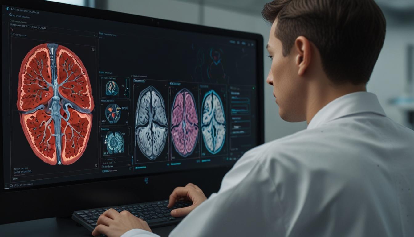
Researchers at the University of California, Los Angeles (UCLA) have made significant strides in medical imaging with the development of a novel deep-learning framework named SLIViT (SLice Integration by Vision Transformer). This system is designed to automatically analyse and diagnose magnetic resonance imaging (MRI) and other 3D medical images, achieving a level of accuracy comparable to that of medical specialists, but within a substantially shorter timeframe.
This pioneering research, detailed in a recent publication in Nature Biomedical Engineering, highlights the adaptability of SLIViT across a myriad of imaging modalities. Initially tested with various types of 3D medical imaging, such as retinal scans, cardiac ultrasound videos, liver MRIs, and chest CT scans, the framework has demonstrated its potential to serve multiple clinical settings through future studies.
3D medical images, due to their volumetric nature, involve more complexity than standard 2D images. Traditional interpretation by clinical specialists requires considerable skill and time, as a single 3D scan consists of numerous 2D slices requiring detailed examination for identifying disease biomarkers. The innovation of SLIViT lies in its ability to circumvent the need for large, manually annotated training datasets, which are often a bottleneck in developing effective AI models for 3D image analysis.
By integrating two artificial intelligence components with an innovative learning strategy, SLIViT effectively leverages existing 2D medical knowledge, enabling it to predict disease risks from comparatively smaller labelled datasets. This efficient process is said to reduce the time taken for analysis by up to 5,000 times without compromising accuracy, as echoed by UCLA PhD student Berkin Durmus, a co-author of the study.
According to Dr. Oren Avram, a postdoctoral researcher and the primary author of the paper, SLIViT's design overcomes the dataset size limitations inherent in traditional models, which typically focus on specific imaging modalities or diseases. By employing a generalised framework, SLIViT shows superior performance across varying 3D image annotation tasks, thereby increasing its clinical applicability by matching the precision of human expertise.
The framework has garnered praise from various experts, including SriniVas R. Sadda, MD, a professor of Ophthalmology at UCLA, who highlighted SLIViT’s proficiency in functioning under practical conditions with limited training samples, which often pose a challenge to typical machine learning models.
Professor Eran Halperin of UCLA underscored the importance of this development, acknowledging the persistent challenge of limited training datasets in clinical environments. SLIViT's capacity to utilise smaller datasets expeditiously allows it to attain performance levels that were previously limited to clinical specialists.
The research team contemplates broadening SLIViT's application, looking into its potential for early disease diagnosis and treatment planning. They also aim to explore strategies to avoid systematic biases in AI, which could lead to health disparities.
In summary, UCLA's SLIViT framework presents a promising advancement in the field of medical imaging. Its ability to automatically and accurately analyse complex 3D medical images with limited data heralds a new era of efficiency in medical diagnostics and research, paving the way for broader clinical applications and more rapid medical advancements.
Source: Noah Wire Services