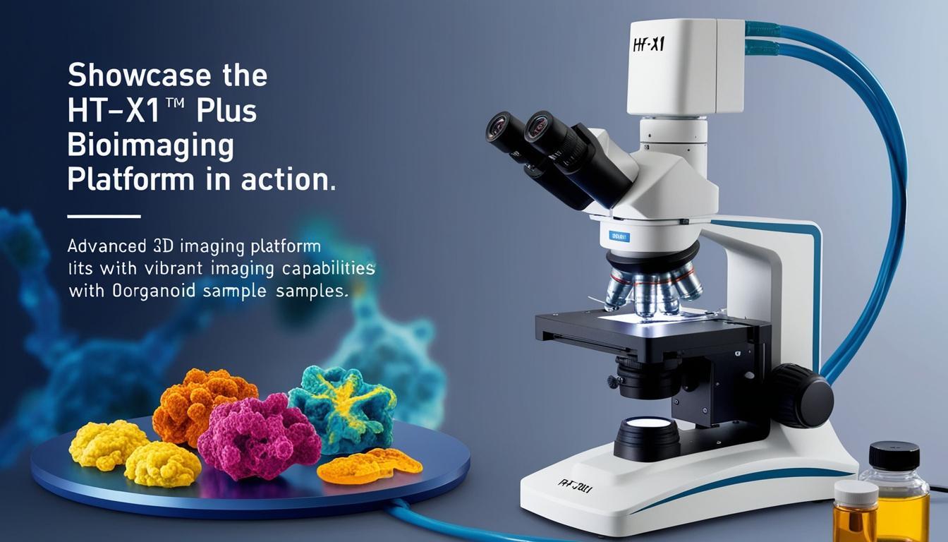
Tomocube, a prominent name in the field of 3D label-free imaging and analysis, has announced the launch of its latest bioimaging platform, the HT-X1™ Plus. This advanced platform is engineered to cater to the critical demands of the biomedical research community, promising significant improvements in high-resolution and high-throughput 3D imaging capabilities for cells and organoids.
The introduction of the HT-X1™ Plus marks a significant leap forward in bioimaging technology. The new platform is equipped with innovative technology enhancements that elevate precision, accelerate imaging speed, and enrich data output. Researchers, who have long been seeking faster and more detailed insights into biological processes, will benefit from the HT-X1™ Plus's ability to offer a field of view that is four times larger, alongside advanced illumination optics. These improvements position the system as a vital tool for phenotypic screening, imaging of tissue sections, and the study of rapid-moving microorganisms. Ultimately, the HT-X1™ Plus opens new avenues for research in areas such as cell biology, regenerative medicine, and studies involving organoids.
Continuing the legacy of the well-regarded HT-X1™ system, the HT-X1™ Plus sets a new benchmark in 3D imaging performance. Integral to its enhancement is a high-performance CXP camera combined with AI-driven image reconstruction algorithms. These features collectively decrease scan times, enabling the comprehensive 3D imaging of an entire 96-well plate in under half an hour without compromising on detail or quality. YongKeun (Paul) Park, the Chief Executive Officer of Tomocube, stated, “With the HT-X1™ Plus, researchers can now explore complex biological samples like organoids with unparalleled clarity and speed. This platform is designed for high-content, image-based drug screening assays and holds tremendous potential to accelerate discoveries in biomedical research.”
Key attributes of the HT-X1™ Plus include an expansive field-of-view, facilitating large-scale experiments without the need for image stitching. The system's rapid image acquisition capabilities make it ideally suited for high-throughput screening, expediting the scanning process for 96-well plates to a brief 30 minutes. Furthermore, the device offers flexible light source configurations using three wavelengths (R/G/B), optimizing contrast and light penetration. The integrated sCMOS-based fluorescence module (FLX™) significantly enhances 3D imaging sensitivity and precision, while the new colour brightfield imaging modality provides improved histological studies and comprehensive tissue morphology analyses.
The HT-X1™ Plus tackles major bioimaging challenges head-on. It addresses issues such as slow scan speeds, limited fluorescence sensitivity, and difficulties with light absorption in dense samples. Its cutting-edge imaging abilities, which minimize phototoxicity and enhance 3D optical sectioning, make it ideally suited for studying dynamic and sensitive samples without losing image quality.
Moreover, the platform's seamless integration with molecular studies and fluorescence-tagged sensors allows researchers to visualise and quantify complex biological processes with exceptional detail. The AI-powered TomoAnalysis™ software offers expansive analysis possibilities, propelling advancements in cellular dynamics research.
Potential applications of the HT-X1™ Plus extend across various domains, including cell biology and regenerative medicine, facilitating high-content screening of live cells and organoids pertinent to drug discovery and therapeutic studies. It also enhances capabilities in 3D biology and organoids research, providing detailed imaging of dense organoids, tissue sections, and immune cell interactions.
In summary, Tomocube's HT-X1™ Plus stands as a pivotal advancement in bioimaging technology, set to empower researchers with improved speed, precision, and data insight, and broaden the frontiers of biomedical discovery.
Source: Noah Wire Services